For US Healthcare
Professionals Only

SEARCH THE SCANS

Look beneath the surface with multimodal imaging

A comprehensive eye examination with imaging can help with early detection, diagnosis, and monitoring of geographic atrophy (GA). Hone in on these signs of early GA in your patients.2,3

Early GA may be difficult to detect

Getting ahead of GA starts with getting to know the biomarkers

See how early GA shows up on different imaging scans.

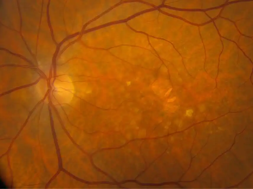
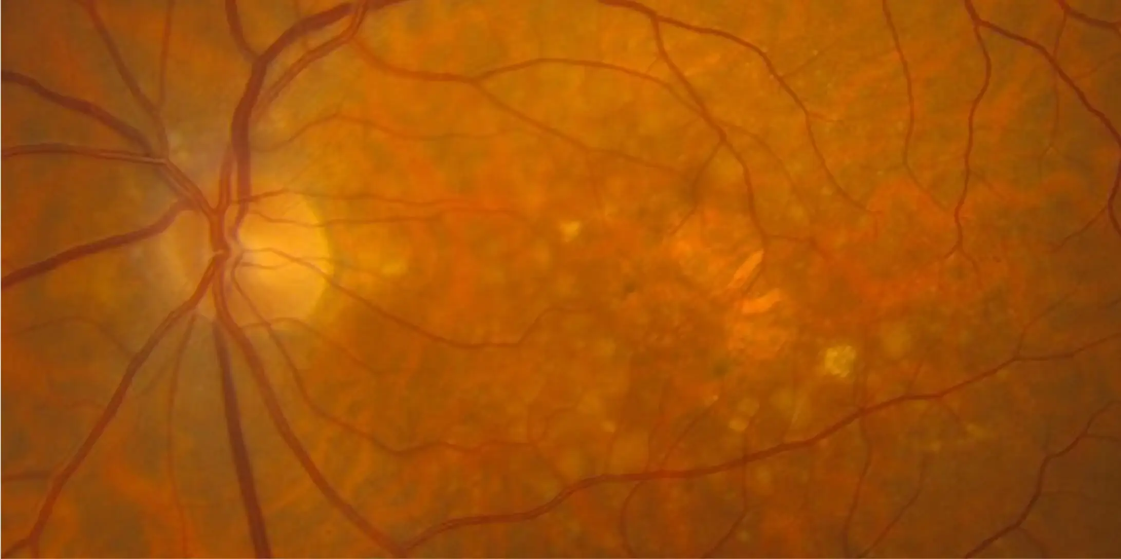
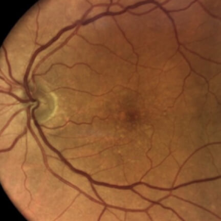
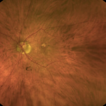
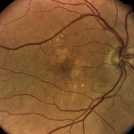
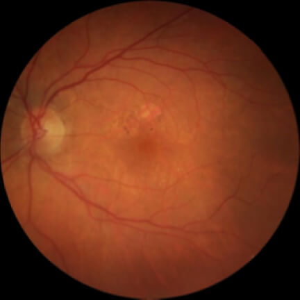
Color Fundus Photography (CFP)

Image courtesy of Dr. Mohammad Rafieetary.

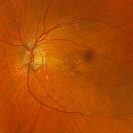
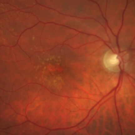
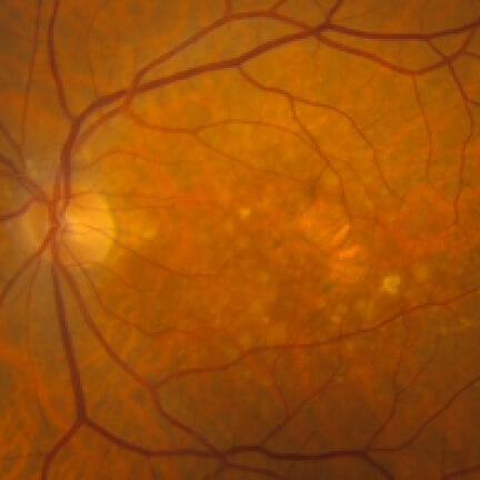
CFPCommonly used for a baseline, although small lesions may be difficult to distinguish3

Identify Biomarkers


Hypopigmented lesions are often circular and sharply demarcated with increased choroidal visibility.2

CFPCommonly used for a baseline, although small lesions may be difficult to distinguish3

Image courtesy of Dr. Mohammad Rafieetary.

Hypopigmented lesions are often circular and sharply demarcated with increased choroidal visibility.2

Identify Biomarkers

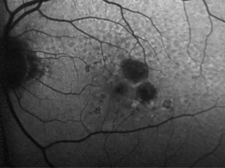
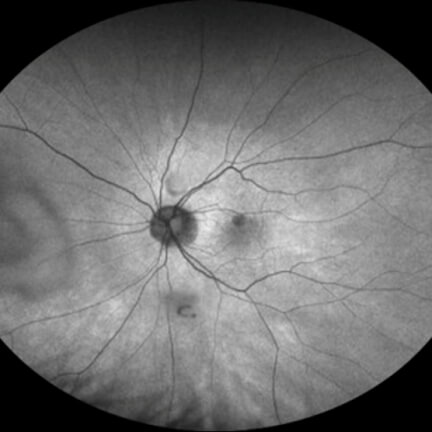
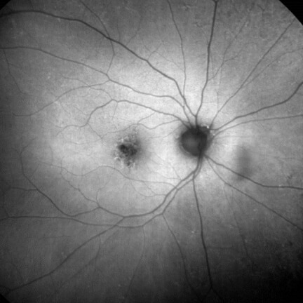
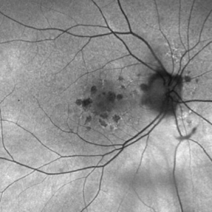
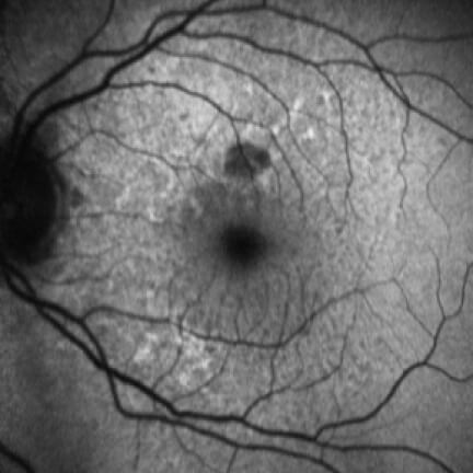
Fundus Autofluorescence (FAF)

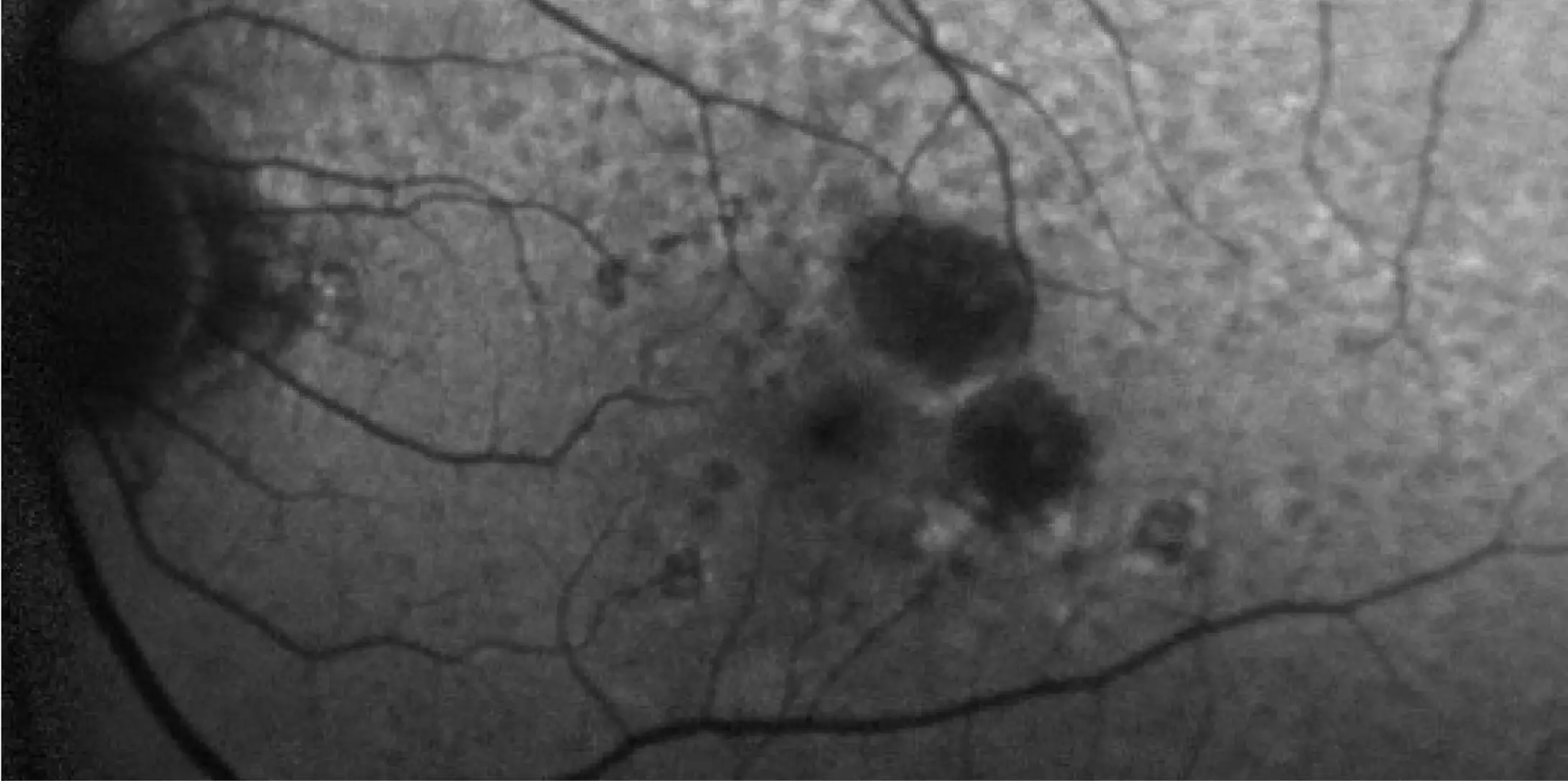
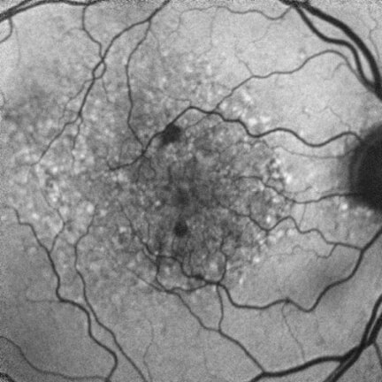
FAFUseful for visualizing progression and educating patients2,3,8

Identify Biomarkers


Dark hypofluorescence indicates areas of atrophic lesions, whereas lighter hyperfluorescent areas highlight accumulation of lipofuscin, which is prognostic of progression.2

FAFUseful for visualizing progression and educating patients2,3,8

Image courtesy of Dr. Julie Rodman.

Dark hypofluorescence indicates areas of atrophic lesions, whereas lighter hyperfluorescent areas highlight accumulation of lipofuscin, which is prognostic of progression.2

Identify Biomarkers

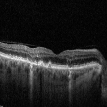
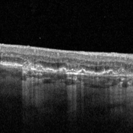
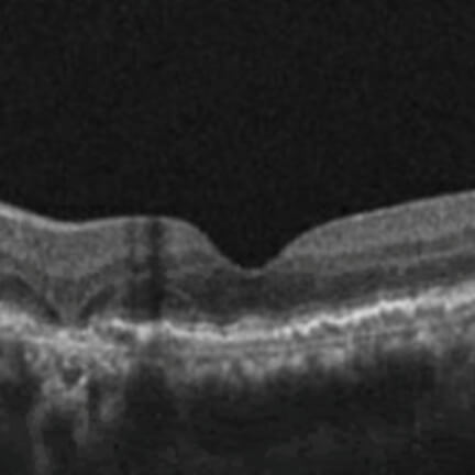
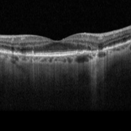
Optical Coherence Tomography (OCT)

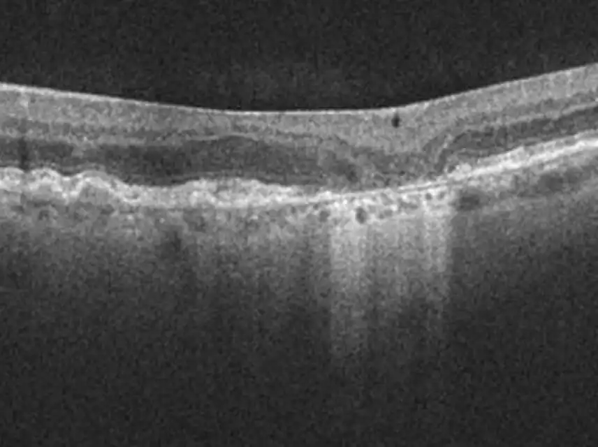
OCTEstablished as a key reference when assessing individuals for initial GA diagnosis8,9

Identify Biomarkers


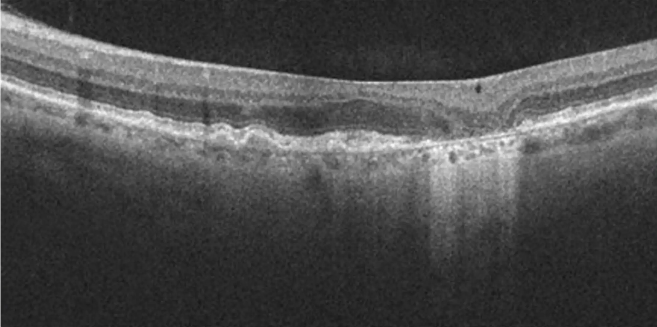
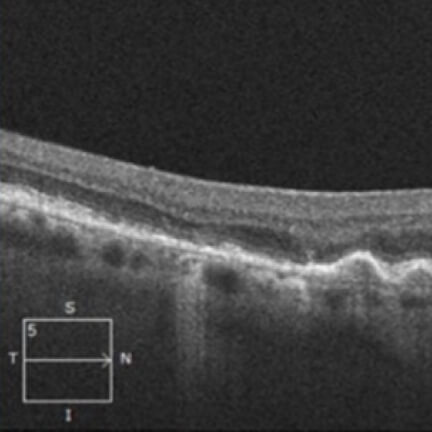
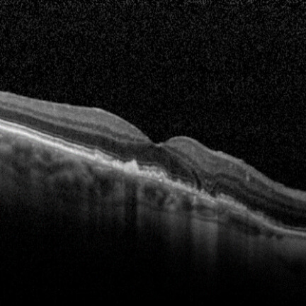
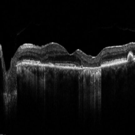
Hypertransmission defects through the choroid point to areas of absent retinal pigment epithelium (RPE), photoreceptors (PR), and choriocapillaris layers.2

OCTEstablished as a key reference when assessing individuals for initial GA diagnosis8,9

Image courtesy of Heidelberg Engineering.

Hypertransmission defects through the choroid point to areas of absent retinal pigment epithelium (RPE), photoreceptors (PR), and choriocapillaris layers.2

Identify Biomarkers

Dr. Julie Rodman

Detecting the signs of early GA

Dr. Julie Rodman explains how multimodal imaging can help you detect GA earlier and assess the risk for progression.

Search the scans and see the signs

See real patient scans from eye care professionals who keep GA on their radar


ASSESS Progression

Be your patient’s advocate to help them slow GA progression with IZERVAY

IMPORTANT SAFETY INFORMATION AND INDICATION

INDICATION

IZERVAY™ (avacincaptad pegol intravitreal solution) is indicated for the treatment of geographic atrophy (GA) secondary to age-related macular degeneration (AMD)

CONTRAINDICATIONS

IZERVAY is contraindicated in patients with ocular or periocular infections and in patients with active intraocular inflammation.

CONTRAINDICATIONS

IZERVAY is contraindicated in patients with ocular or periocular infections and in patients with active intraocular inflammation.

WARNINGS AND PRECAUTIONS

Endophthalmitis and Retinal Detachments

  • Intravitreal injections, including those with IZERVAY, may be associated with endophthalmitis and retinal detachments. Proper aseptic injection technique must always be used when administering IZERVAY in order to minimize the risk of endophthalmitis. Patients should be instructed to report any symptoms suggestive of endophthalmitis or retinal detachment without delay and should be managed appropriately.

Neovascular AMD

  • In clinical trials, use of IZERVAY was associated with increased rates of neovascular (wet) AMD or choroidal neovascularization (7% when administered monthly and 4% in the sham group) by Month 12. Patients receiving IZERVAY should be monitored for signs of neovascular AMD.

Increase in Intraocular Pressure

  • Transient increases in intraocular pressure (IOP) may occur after any intravitreal injection, including with IZERVAY. Perfusion of the optic nerve head should be monitored following the injection and managed appropriately.

ADVERSE REACTIONS

Most common adverse reactions (incidence ≥5%) reported in patients receiving IZERVAY were conjunctival hemorrhage, increased IOP, blurred vision, and neovascular age-related macular degeneration.

INDICATION

IZERVAY™ (avacincaptad pegol intravitreal solution) is indicated for the treatment of geographic atrophy (GA) secondary to age-related macular degeneration (AMD)

Please see full Prescribing Information.

IMPORTANT SAFETY INFORMATION AND INDICATION

CONTRAINDICATIONS

IZERVAY is contraindicated in patients with ocular or periocular infections and in patients with active intraocular inflammation.

WARNINGS AND PRECAUTIONS

Endophthalmitis and Retinal Detachments

  • Intravitreal injections, including those with IZERVAY, may be associated with endophthalmitis and retinal detachments. Proper aseptic injection technique must always be used when administering IZERVAY in order to minimize the risk of endophthalmitis. Patients should be instructed to report any symptoms suggestive of endophthalmitis or retinal detachment without delay and should be managed appropriately.

Neovascular AMD

  • In clinical trials, use of IZERVAY was associated with increased rates of neovascular (wet) AMD or choroidal neovascularization (7% when administered monthly and 4% in the sham group) by Month 12. Patients receiving IZERVAY should be monitored for signs of neovascular AMD.

Increase in Intraocular Pressure

  • Transient increases in intraocular pressure (IOP) may occur after any intravitreal injection, including with IZERVAY. Perfusion of the optic nerve head should be monitored following the injection and managed appropriately.

ADVERSE REACTIONS

Most common adverse reactions (incidence ≥5%) reported in patients receiving IZERVAY were conjunctival hemorrhage, increased IOP, blurred vision, and neovascular age-related macular degeneration.

INDICATION

IZERVAY™ (avacincaptad pegol intravitreal solution) is indicated for the treatment of geographic atrophy (GA) secondary to age-related macular degeneration (AMD)

Please see full Prescribing Information.

Images courtesy of Heidelberg Engineering, Dr. Julie Rodman, and Dr. Mohammad Rafieetary.