For US Healthcare
Professionals Only

Assess Progression

Keep the risks of rapid GA progression on your radar

With geographic atrophy (GA), time is of the essence.2,3 Search for the signs of rapid progression to identify which of your patients are most at risk.

GA progression is highly variable

Look for the risk factors prognostic of rapid progression

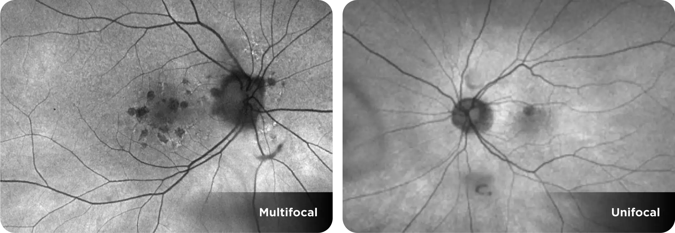
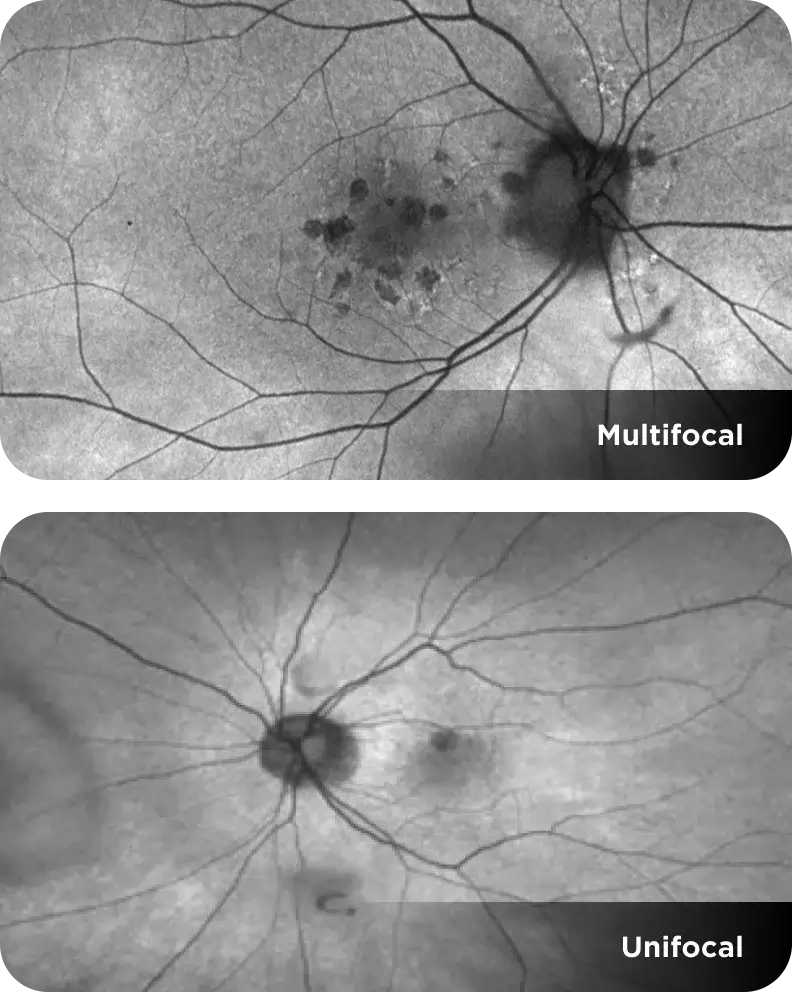
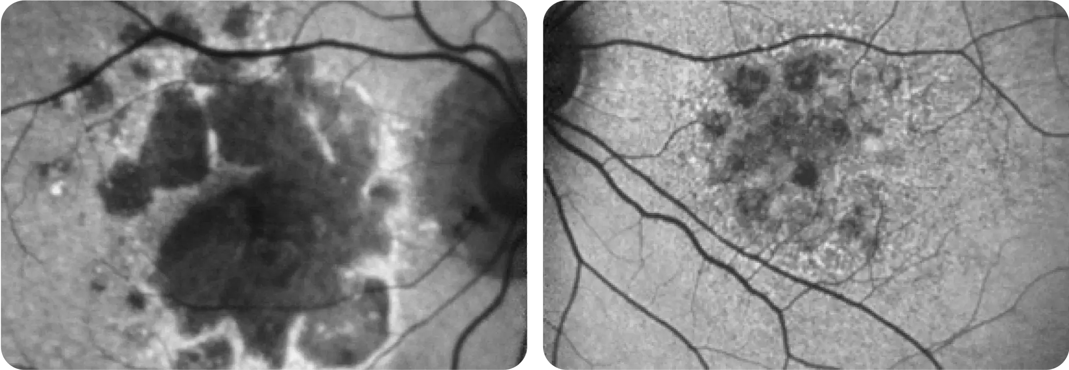
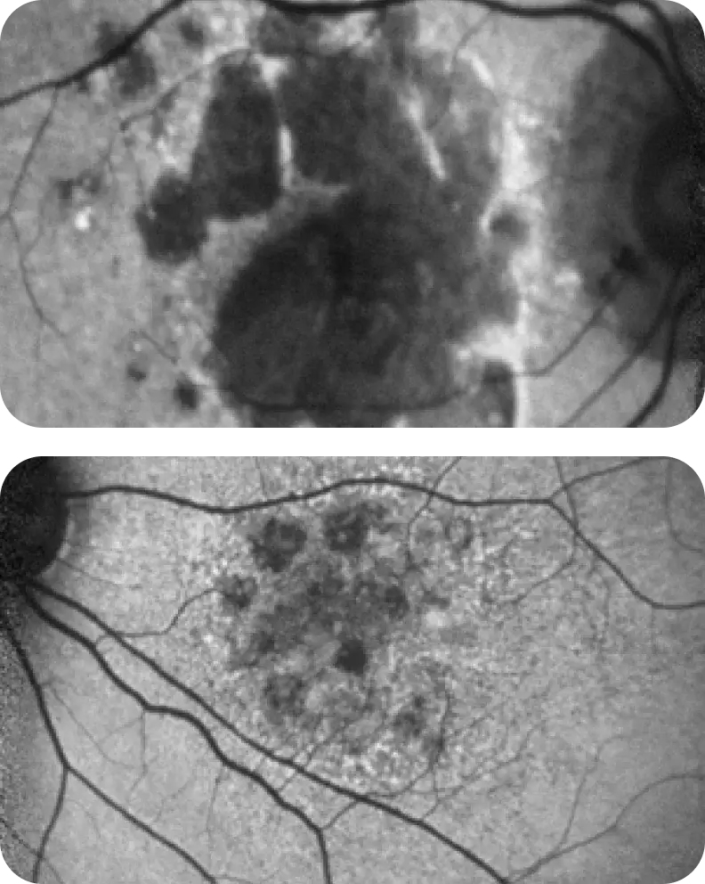
Focality

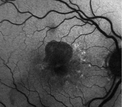
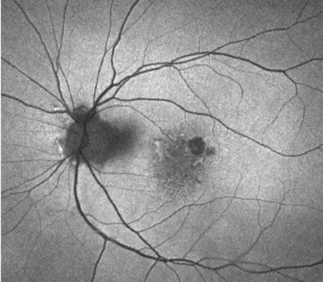
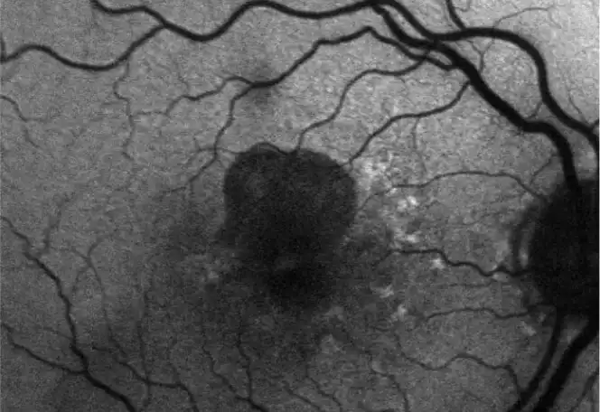
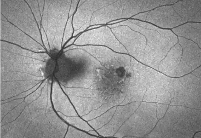
Multifocal lesions often grow faster than unifocal lesions.2
Images courtesy of Dr. Julie Rodman ("Unifocal") and Dr. Mohammad Rafieetary ("Multifocal").

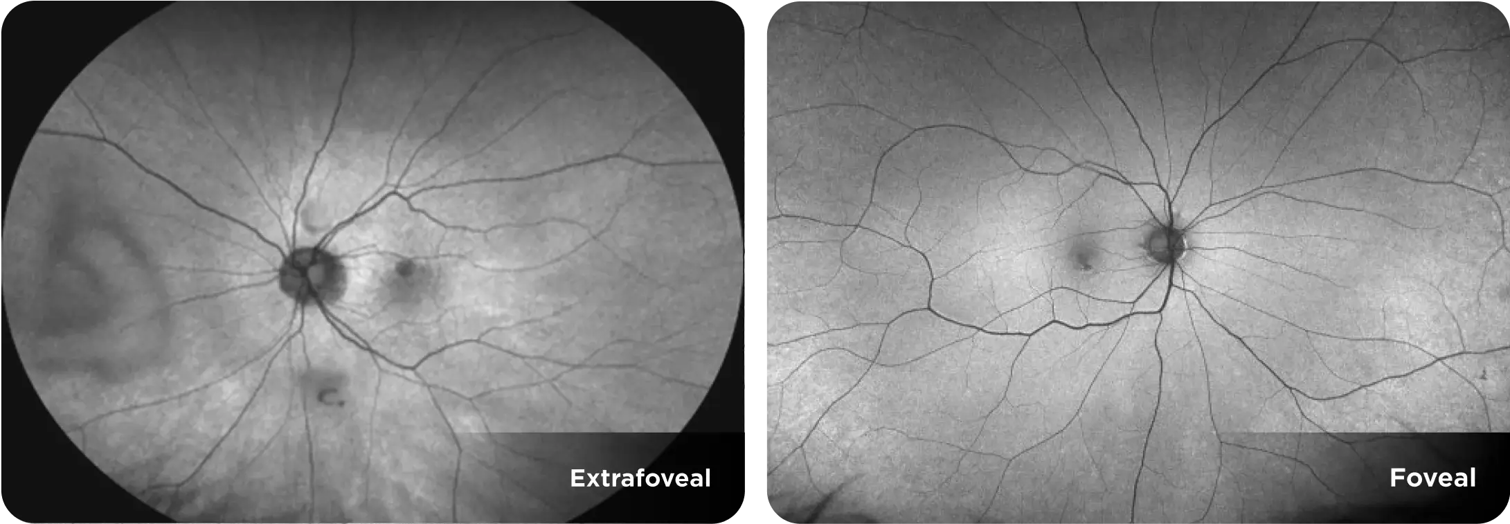
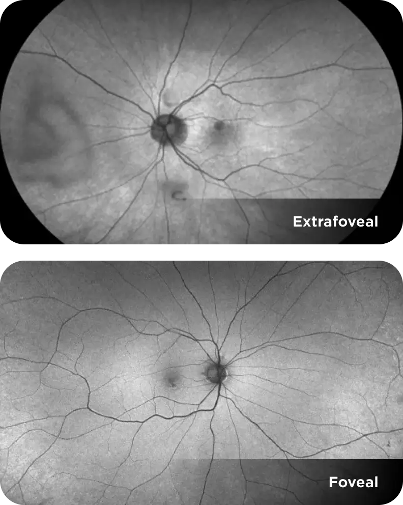
Location

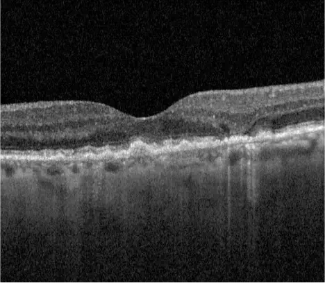
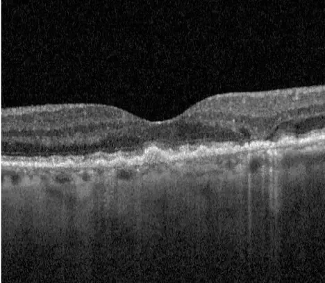
Extrafoveal lesions often grow faster than foveal lesions.2
Images courtesy of Heidelberg Engineering ("Foveal") and Dr. Julie Rodman (“Extrafoveal").

Hyperfluorescence

Hyperfluorescence highlights areas at risk for progression as a result of lipofuscin accumulation.2,3
Images courtesy of Dr. Mary Beth Yackey.

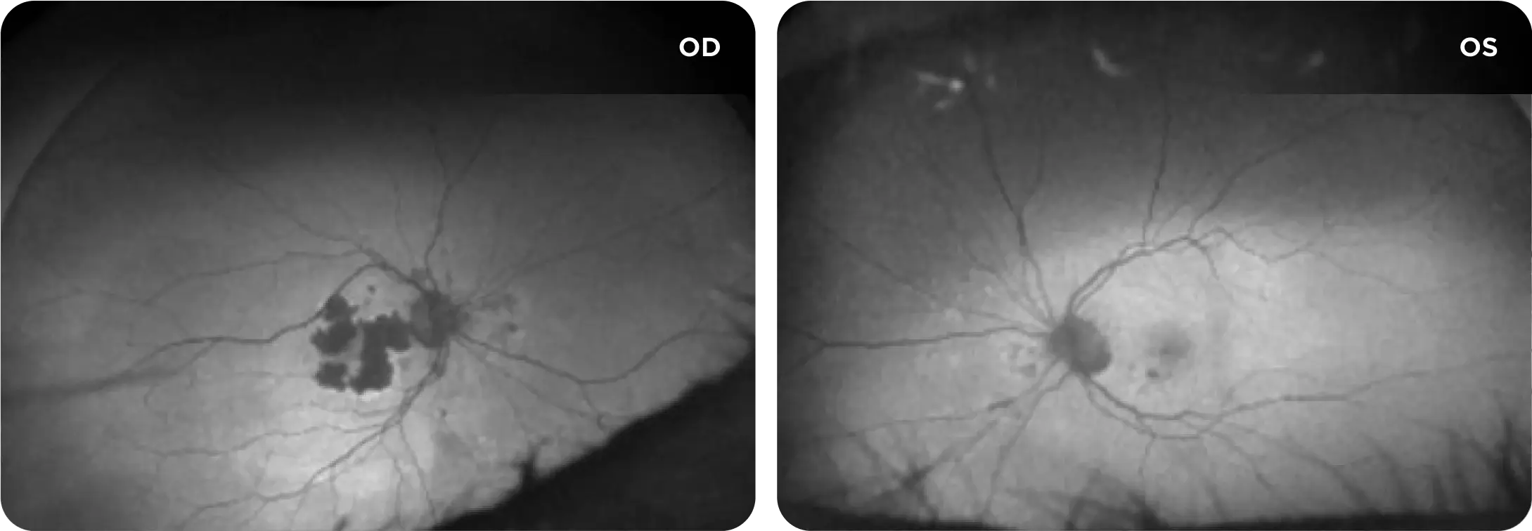
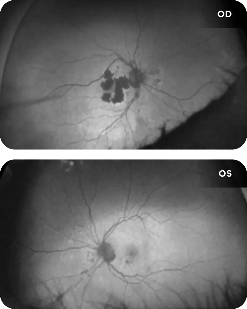
Laterality

GA often develops bilaterally, at which point it may grow faster.2
Images courtesy of Dr. Julie Rodman.

Assess the risks to get ahead of GA progression

The following cases feature real scans from patients with GA at risk of rapid progression if left untreated.*

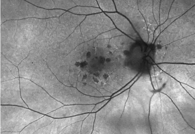
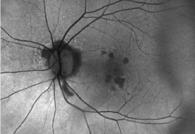
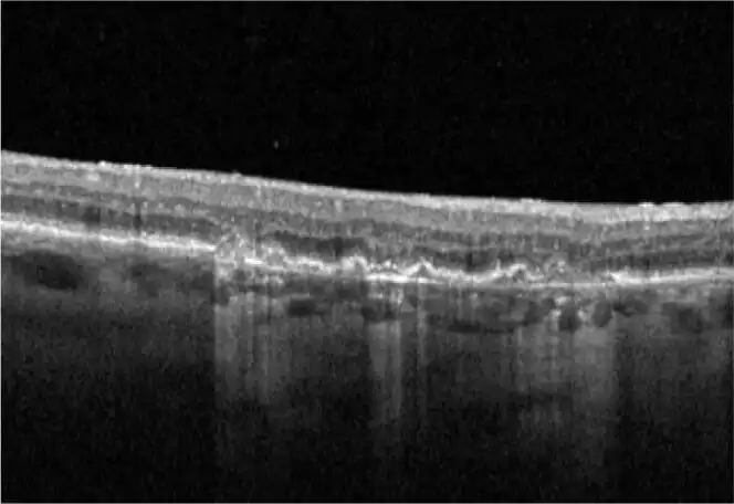
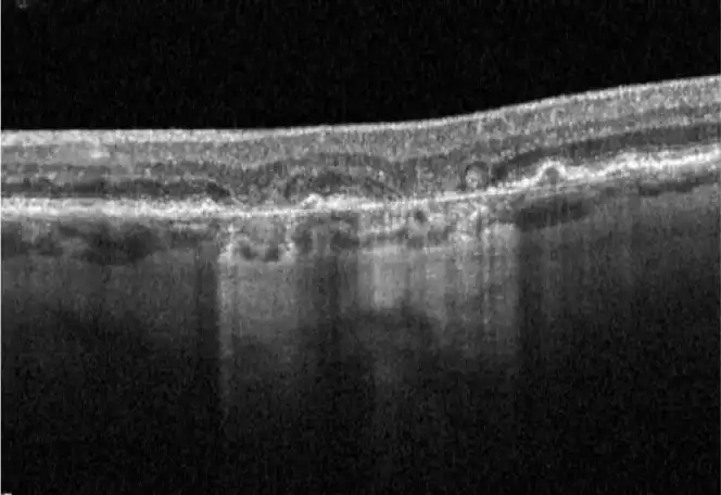
This patient has good visual acuity, but multifocal GA threatens the fovea on multiple fronts.

Clinical History
  • 63-year-old White female
  • BCVA OS declined from 20/25-2 to 20/30
  • Patient complains of difficulty reading at night
Additional Factors
  • Mother had AMD

Risk Assessment Summary

Although this patient has relatively good visual acuity at this point, it’s important to note that BCVA does not necessarily correlate with visual function.1,2

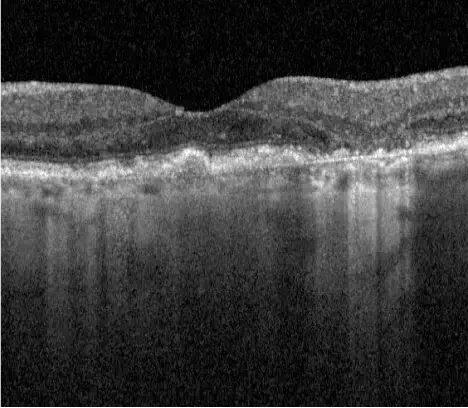
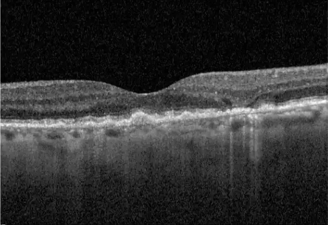
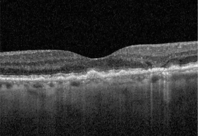
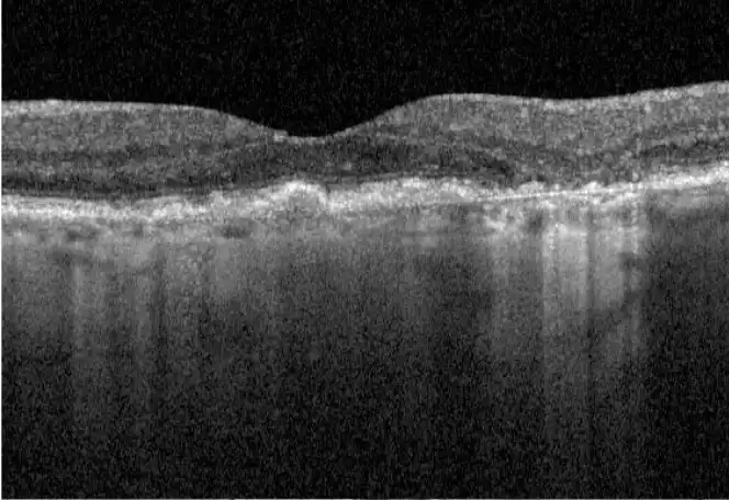
  • The multifocal, extrafoveal lesions are easily visualized on FAF and OCT
  • On FAF (specifically OD), a diffuse hyperfluorescent pattern can be seen
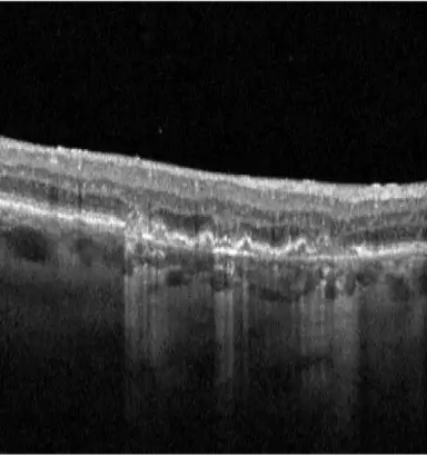
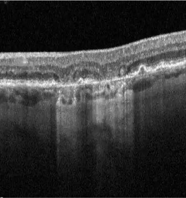
  • On OCT, multiple bands of choroidal hypertransmission and RPE and PR layer loss can also be seen

The presence of bilateral, multifocal, extrafoveal lesions and hyperfluorescent areas coupled with a family history of AMD increases this patient’s risk of rapid progression.2

An early referral for IZERVAY may help slow the impending irreversible effects of GA.2-4

AMD=age-related macular degeneration.

*These case studies feature real scans, but some historical details have been modified for educational purposes.

This patient’s progressing GA puts their remaining visual function at risk.2

Clinical History
  • 75-year-old Asian female
  • Cataract surgery with PCIOL OS 20/40, OD 20/400
  • Expresses concern about losing vision in left eye
Additional Factors
  • COPD

Risk Assessment Summary

Given that GA has already significantly affected visual acuity in the right eye, it is imperative to stay vigilant for risk factors in the fellow eye.2

  • This patient has multifocal lesions and hyperfluorescent areas exhibited on FAF bilaterally
  • Additionally, OCT (OS) shows an increase in size of choroidal hypertransmission over a 3-year period

The presence of multifocal lesions, hyperfluorescent areas, and previous rate of progression elevates this patient’s risk of further progression in both eyes.2

This patient is in urgent need of a referral to a retinal specialist who may recommend IZERVAY to help slow the GA progression.4

COPD=chronic obstructive pulmonary disease; PCIOL=posterior chamber intraocular lens.

*These case studies feature real scans, but some historical details have been modified for educational purposes.

This patient’s lesions are critically close, with early GA already encroaching on the fovea.

Clinical History
  • 69-year-old Hispanic male
  • Visual acuity 20/25 OD, 20/20 OS
  • Complains of blurry vision and difficulty driving at night
  • OS has intermediate AMD
Additional Factors
  • Smoker for 25 years
  • Hypertension

Search the scans

Risk Assessment Summary

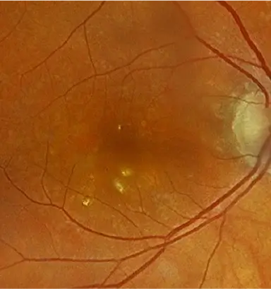
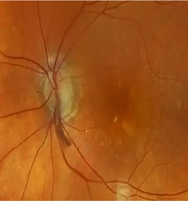
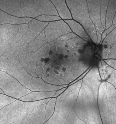
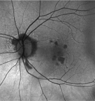
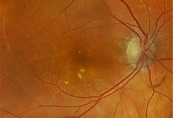
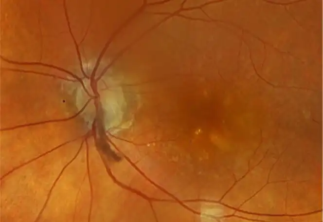
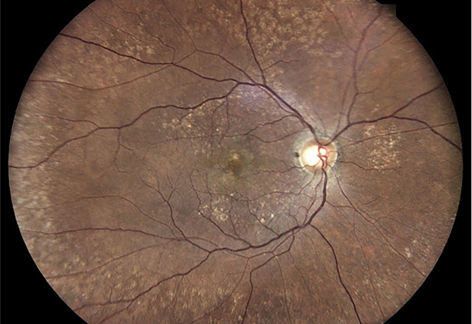
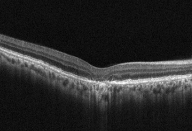
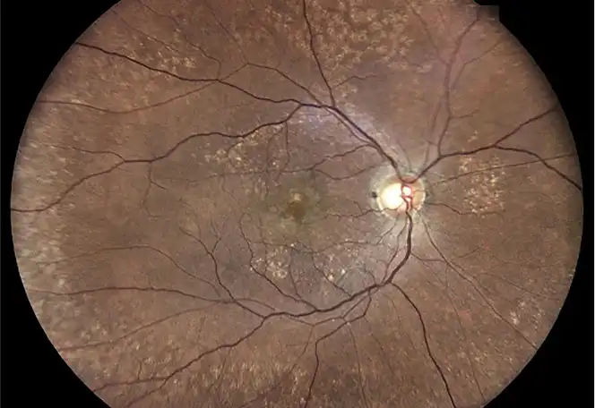
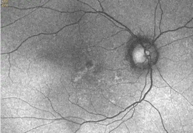
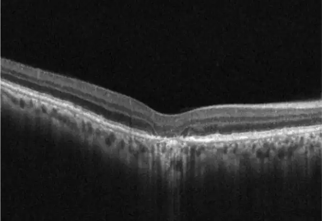
The signs of this patient’s GA on CFP are subtle, but they are more apparent on FAF and OCT.

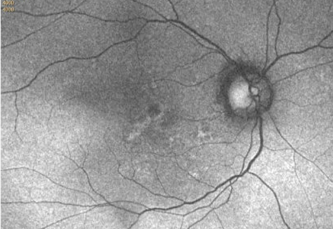
  • This patient’s FAF shows multifocal lesions surrounded by hyperfluorescent areas
  • Additionally, choroidal hypertransmission on OCT shows GA encroaching on the fovea, with little latitude until central foveal involvement

Closer inspection of these images confirms risk factors linked to rapid GA progression, including multifocality, hyperfluorescent areas, and an extrafoveal lesion.2

This patient’s timely diagnosis optimizes the opportunity to get ahead of GA progression.2-4

AMD=age-related macular degeneration.

*These case studies feature real scans, but some historical details have been modified for educational purposes.

Download Case Studies

Act Early

A prompt referral for IZERVAY can make a significant difference4

IMPORTANT SAFETY INFORMATION AND INDICATION

INDICATION

IZERVAY™ (avacincaptad pegol intravitreal solution) is indicated for the treatment of geographic atrophy (GA) secondary to age-related macular degeneration (AMD)

CONTRAINDICATIONS

IZERVAY is contraindicated in patients with ocular or periocular infections and in patients with active intraocular inflammation.

CONTRAINDICATIONS

IZERVAY is contraindicated in patients with ocular or periocular infections and in patients with active intraocular inflammation.

WARNINGS AND PRECAUTIONS

Endophthalmitis and Retinal Detachments

  • Intravitreal injections, including those with IZERVAY, may be associated with endophthalmitis and retinal detachments. Proper aseptic injection technique must always be used when administering IZERVAY in order to minimize the risk of endophthalmitis. Patients should be instructed to report any symptoms suggestive of endophthalmitis or retinal detachment without delay and should be managed appropriately.

Neovascular AMD

  • In clinical trials, use of IZERVAY was associated with increased rates of neovascular (wet) AMD or choroidal neovascularization (7% when administered monthly and 4% in the sham group) by Month 12. Patients receiving IZERVAY should be monitored for signs of neovascular AMD.

Increase in Intraocular Pressure

  • Transient increases in intraocular pressure (IOP) may occur after any intravitreal injection, including with IZERVAY. Perfusion of the optic nerve head should be monitored following the injection and managed appropriately.

ADVERSE REACTIONS

Most common adverse reactions (incidence ≥5%) reported in patients receiving IZERVAY were conjunctival hemorrhage, increased IOP, blurred vision, and neovascular age-related macular degeneration.

INDICATION

IZERVAY™ (avacincaptad pegol intravitreal solution) is indicated for the treatment of geographic atrophy (GA) secondary to age-related macular degeneration (AMD)

Please see full Prescribing Information.

IMPORTANT SAFETY INFORMATION AND INDICATION

CONTRAINDICATIONS

IZERVAY is contraindicated in patients with ocular or periocular infections and in patients with active intraocular inflammation.

WARNINGS AND PRECAUTIONS

Endophthalmitis and Retinal Detachments

  • Intravitreal injections, including those with IZERVAY, may be associated with endophthalmitis and retinal detachments. Proper aseptic injection technique must always be used when administering IZERVAY in order to minimize the risk of endophthalmitis. Patients should be instructed to report any symptoms suggestive of endophthalmitis or retinal detachment without delay and should be managed appropriately.

Neovascular AMD

  • In clinical trials, use of IZERVAY was associated with increased rates of neovascular (wet) AMD or choroidal neovascularization (7% when administered monthly and 4% in the sham group) by Month 12. Patients receiving IZERVAY should be monitored for signs of neovascular AMD.

Increase in Intraocular Pressure

  • Transient increases in intraocular pressure (IOP) may occur after any intravitreal injection, including with IZERVAY. Perfusion of the optic nerve head should be monitored following the injection and managed appropriately.

ADVERSE REACTIONS

Most common adverse reactions (incidence ≥5%) reported in patients receiving IZERVAY were conjunctival hemorrhage, increased IOP, blurred vision, and neovascular age-related macular degeneration.

INDICATION

IZERVAY™ (avacincaptad pegol intravitreal solution) is indicated for the treatment of geographic atrophy (GA) secondary to age-related macular degeneration (AMD)

Please see full Prescribing Information.

Images courtesy of Heidelberg Engineering, Dr. Julie Rodman, and Dr. Mohammad Rafieetary.